


It is a period of hydraulic confusion. The venous reservoir appears to supply the force returning blood to the heart, while the cardiac pump merely restores what has been lost. But an old law of physics has been disturbed. A reservoir that never empties may have no energy left to give…
The appeal of Magder’s bathtub was easy to understand.
The water stored in the tub represented blood held within the compliant venous system. Its depth represented mean systemic filling pressure, Pms. The drain led towards the right atrium, with the difference between Pms and right atrial pressure providing the pressure head for venous return. The heart appeared as a tap, replacing the water that escaped and preserving the state of the reservoir.
Most of the ingredients were real. The veins contain most of the circulating blood. Their walls are elastic. Blood volume distends them and creates stored energy. Open a large vein to atmospheric pressure and blood continues to leave after the heart has stopped, propelled for a time by vascular recoil.
Magder accepted that this flow could not continue indefinitely. As the vascular system emptied, its pressure would fall and the stored energy would be exhausted. The heart was therefore needed to return blood to the reservoir and restore the energy released during drainage.
This gave the heart a role, but a limited one. The venous reservoir determined how much blood could drain. The heart permitted that flow by keeping right atrial pressure low and restored the volume that had been lost.
Brengelmann saw a problem hidden inside this division of labour.
If the heart continuously restored everything leaving the reservoir, the reservoir would remain at the same average volume and pressure. Its elastic walls would remain equally distended.
How, then, could they continuously release energy?
No recoil without emptying
An elastic structure releases stored energy by changing shape.
A stretched spring releases energy as it shortens. An inflated balloon releases energy as it becomes smaller. A distended vein can release energy as its contained volume falls and its wall recoils.
This is exactly what happens when the circulation stops and a vein is opened to atmospheric pressure. The vascular system has been connected to a new, lower-pressure boundary. Blood leaves. Vascular volume falls. The walls recoil and the elastic energy stored within them is released.
The flow is real, but it is transient. As the vessels empty, their pressure falls. Eventually the available energy has been spent and flow stops.
Steady venous return is different. Blood may be flowing rapidly through a vascular compartment while the amount of blood contained within it remains unchanged. If five litres enter each minute and five litres leave, the compartment passes a flow of five litres per minute without losing any volume.
Throughput is not the same as emptying.
In the steady state, average inflow and outflow are equal. The compartment does not become progressively smaller and its walls do not progressively recoil. Blood passes through it, but its stored elastic energy remains stored.
This was the meaning of Brengelmann’s terse statement that there could be “no energy release” without a decrease in volume. The mathematics expresses mechanical work as pressure acting through a change in volume. If the volume does not fall, the elastic wall has not performed net work on the blood.
Magder’s reservoir was continuously drained, but it was also continuously refilled. Something therefore had to return each lost volume element and restore its energy. In the bathtub this work was hidden inside the tap. In the circulation it was performed principally by the heart.
The reservoir still mattered. Its volume and elastic properties determined its pressure state and influenced how the circulation responded to a change in cardiac activity. What it could not do was remain unchanged while also acting as a continuous source of mechanical work.
The physics belong to the bathtub
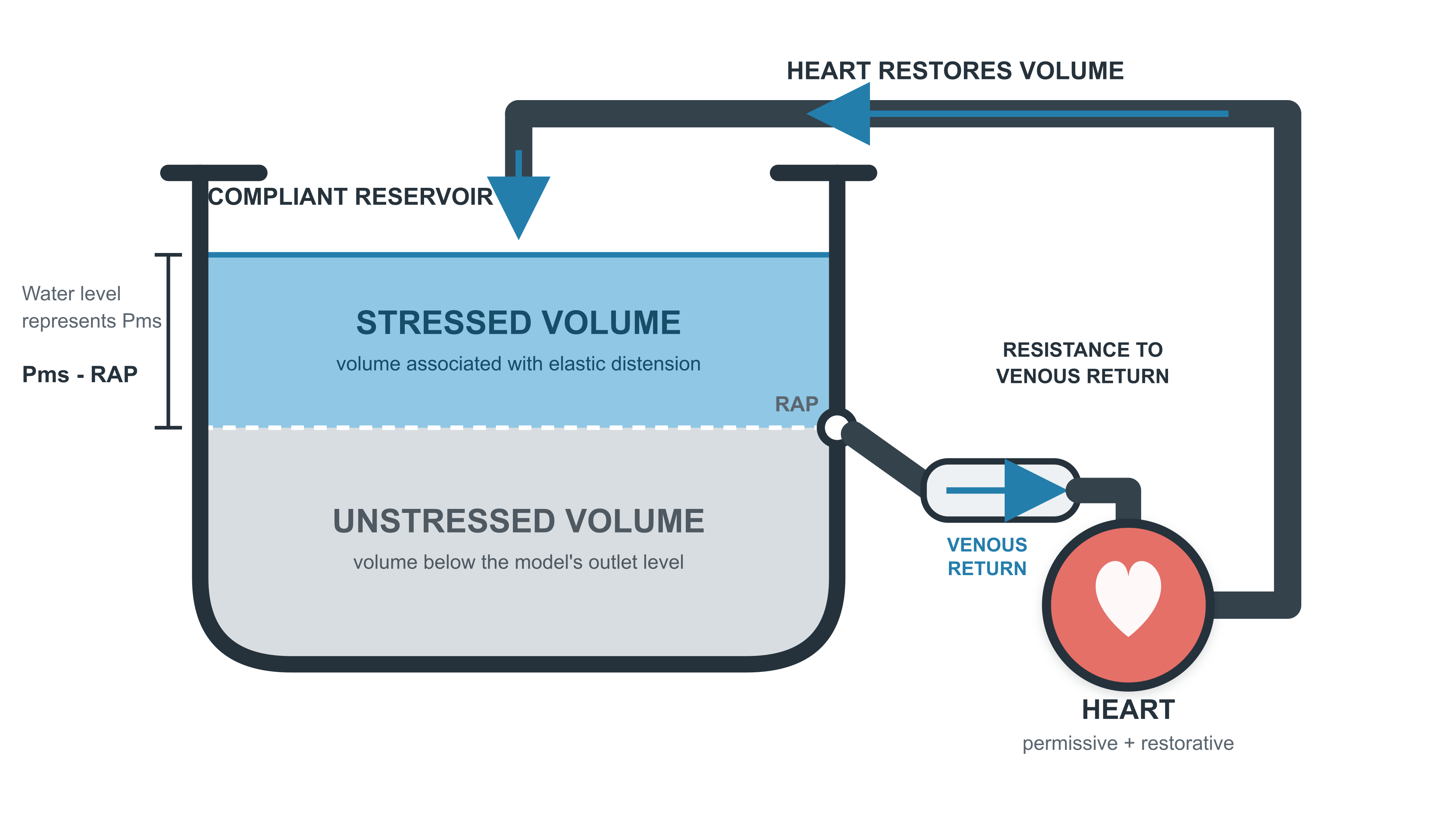
The bathtub analogy feels natural because its source of energy is visible.
Water in the tub sits above the drain. Gravity acts on it continuously. As water descends, gravitational potential energy is converted into movement and then dissipated by resistance in the outlet.
If the water level is to remain constant, the tap must replace what leaves. But that replacement water must first be returned to the height of the tub. Somewhere outside the picture, a pump, elevated reservoir or pressurised water supply has performed the work needed to restore its gravitational potential energy.
The tap is therefore doing more than keeping an inventory. It is restoring the energy that gravity releases through the drain.
In reality there is no equivalent independent gravitational reservoir in the circulation. Blood does not travel around the systemic circulation because its venous end sits physically above the heart. The heart supplies mechanical energy to a closed vascular system. Its work, together with vascular resistance, compliance and blood volume, establishes the flowing distribution of pressure and volume.
The bathtub gives the venous reservoir a separate gravitational energy source that the circulation does not possess.
Its geometry creates another problem. In the illustrations of high venous return, the heart is placed below the outlet of the tub. Lowering it would increase the hydrostatic pressure measured at heart level. Yet the Guyton curve associates higher flow with a lower right atrial pressure. Correcting all pressures to the same vertical reference would expose the mismatch.
The arterial side has largely disappeared as well. The tap pours cardiac output back into the tub as though it arrives at atmospheric pressure. In the circulation, the left ventricle ejects into a high-pressure arterial system. Blood then passes through a succession of real vascular segments, each with its own resistance, compliance, pressure and contained volume.
The analogy makes another point about the heart.
In Magder’s model, the heart lowers right atrial pressure and replaces the blood that drains from the reservoir. If it is too weak to do this, it limits flow. Once it is no longer the limiting element, however, greater cardiac power cannot make the reservoir drain faster. Flow is then set by the stressed volume and drainage characteristics of the vascular system. The heart permits and restores that flow, but does not determine its magnitude.
Magder supports this by pointing out that the heart contains very little blood compared with the veins and venules. It cannot add much volume to the venous reservoir from its own contents or rapidly increase the reservoir’s elastic pressure.
This captures an important truth. Steady flow depends on what the systemic circulation can deliver and what the heart can accept and eject. If cardiac function is limiting, improving it can increase flow. Once the heart can readily handle the blood presented to it, additional power has little effect unless the vascular state also changes.
The heart’s small contained volume is relevant, but it is not the whole explanation. A pump influences a circuit through the volume it moves over time as well as the volume held inside it at one moment.
The heart continually transfers blood between vascular compartments. This helps establish how much blood resides on the arterial and venous sides, and therefore the pressures observed during flow. It can alter this distribution even when total blood volume, vascular accommodation and true zero-flow Pms remain unchanged.
The bathtub therefore captures a real vascular constraint. Once the heart is no longer limiting, the vascular state restricts how much flow can be sustained. What the analogy does not establish is that the venous reservoir supplies the continuous energy for that flow. The heart provides the energy, while the vascular state constrains the flow that results.
An aggregate without an address
Defenders of the Guytonian account do not all require a single anatomical reservoir maintained at Pms.
Berger, Moller and Takala describe Pms as the aggregate pressure of the entire systemic vasculature. It represents systemic stressed volume acting within the combined compliance of many vascular beds. No single compartment needs to sit at precisely that pressure during flow.
This is a more sophisticated account than the bathtub. It preserves the importance of the vascular elastic state without claiming that Pms must reside in one particular vein.
It also creates a problem for the original gradient.
A spatial pressure difference exists between two locations at the same time. Blood flowing through a vein encounters one local pressure at its upstream end and another at its downstream end. Those pressures and the flow develop together within the operating circulation.
Pms has a different origin. It is the common pressure approached when systemic flow stops and blood redistributes between vascular compartments. If it is instead treated as a weighted description of the whole system during flow, it still lacks a single upstream location from which blood travels to the right atrium.
Magder’s more recent account takes a different route. He distinguishes mean circulatory filling pressure, the equilibrium pressure of the whole circulation at zero flow, from mean systemic filling pressure during flow. Because the systemic veins are so compliant, their pressure changes relatively little, allowing him to place the flowing value of mean systemic filling pressure within that region and retain it as the upstream pressure for venous return.
A local systemic venous pressure is physically real. It can form one end of a real pressure difference across the vascular segments downstream from it. But it is also determined by the volume residing in that region, its compliance, the surrounding pressure and the flow through the connected circulation. It cannot simply inherit the identity of the equilibrium pressure measured after flow stops.
Numerical similarity is not physical identity.
The vascular elastic state is real. Local venous pressures are real. Neither fact demonstrates that the zero-flow pressure Pms persists at an anatomical location during flow and continuously supplies the energy for venous return.
The curve without a reservoir
Brengelmann’s 2019 analysis offered another explanation for the famous venous return curve.
Begin with the circulation at zero flow. Pressures have equilibrated at Pms. Blood is distributed among the arterial and venous compartments according to their volume-containing properties.
Now start the pump.
The pump initially transfers blood into the arterial compartments faster than it leaves them. Their contained volume rises, their walls distend and arterial pressure rises above Pms. Because total blood volume is fixed, this additional arterial volume comes from the venous side. The venous compartments contain less blood and their pressures fall below Pms.
Right atrial pressure falls as part of this redistribution.
Once the redistribution is complete, inflow and outflow again match in every compartment. Flow continues through the new pressure profile while the compartments remain at their new volumes.
Increase pump flow again and the redistribution becomes larger. More volume resides on the arterial side and less on the venous side. Arterial pressure rises further while right atrial pressure falls further. Repeating the process produces the familiar inverse relationship between flow and right atrial pressure.
No compartment has remained at Pms. No reservoir at Pms has drained through a single venous resistance. The curve has emerged from flow, vascular resistance, compliance and conservation of blood volume.
Real pressure differences exist throughout this model. Each connects two neighbouring locations in the flowing circulation. The problem lies specifically with treating Pms and RAP as if they were the two ends of one physical pathway.
Guyton’s original mathematics had described the redistribution correctly. The result could be written so that flow was proportional to Pms minus right atrial pressure. But Brengelmann showed that the same volume accounting could instead be written from the arterial end, making flow proportional to arterial pressure minus Pms.
If the position of a term within an equation established causality, this second arrangement would invite an equally strange conclusion: Pms was a back pressure opposing arterial delivery, and cardiac output could be increased by lowering it.
Neither reading follows from the mathematics.
The denominator traditionally called resistance to venous return creates a similar illusion. It sounds like the resistance of the veins between an upstream Pms reservoir and the right atrium. In Guyton’s model, however, its value depended on arterial as well as venous resistance and on the distribution of compliance across the network. Guyton initially called it an impedance to venous return, although it was not impedance in the usual frequency-dependent sense.
It was an effective parameter describing the behaviour of the whole vascular system, not an anatomical resistor placed downstream from Pms.
The equation was performing vascular bookkeeping. It related flow to the pressure and volume distribution that accompanied it. It did not identify a hidden motor at Pms or a discrete resistance lying between Pms and the right atrium.
What PEEP reveals
Positive end-expiratory pressure provides a useful test of the supposed driving gradient. It raises pressure around the heart and thoracic veins, alters cardiac filling and redistributes blood between the pulmonary, arterial and systemic venous compartments.
Jellinek and colleagues studied patients undergoing testing of implanted defibrillators. Before cardiac arrest, average right atrial pressure was 7.3 mmHg. The systemic venous pressure measured after flow stopped was 10.2 mmHg, giving a Pms–RAP difference of approximately 3 mmHg.
When PEEP was increased from 0 to 15 cmH₂O, right atrial pressure rose to 10.0 mmHg and Pms rose to 12.7 mmHg. The difference between them was almost unchanged.
Stroke volume nevertheless fell by approximately 23%.
The equation could accommodate this result by assigning the fall in flow to an increase in resistance to venous return. But that calculation cannot explain what caused the new state. The central observation remains: substantially different flows were associated with the same Pms–RAP difference. The gradient did not independently determine flow.
Berger and colleagues later demonstrated similar complexity in pigs. Raising PEEP increased both right atrial pressure and directly measured stop-flow Pms. The increase in PEEP was much smaller than in Jellinek’s study, and blood flow fell only slightly. During inspiratory holds, flow from different venous regions fell and recovered at different rates. The systemic veins behaved as a network of compliant compartments that exchanged volume and responded differently, rather than as a single reservoir draining uniformly towards the heart.
PEEP changed cardiac function and the distribution of blood around the circulation. More blood could reside in the systemic veins while less remained in the pulmonary and arterial compartments. The resulting volume changes were expressed as changes in pressure. Pms and right atrial pressure could therefore rise together even while cardiac output fell.
These experiments did not invalidate the mathematical relationship between Pms, right atrial pressure, flow and resistance. They showed its proper status. The gradient described the pressure distribution accompanying the new circulatory state; it did not determine the flow on its own.
Can pulsatility save the reservoir?
The real circulation is not perfectly steady.
Within each heartbeat, inflow and outflow from individual vascular compartments do not match at every instant. Their volumes fluctuate. Elastic walls expand, recoil and exchange energy with the blood.
Berger and colleagues argued that this pulsatility matters. Stressed volume does not disappear when the circulation is flowing. Small changes in venous volume allow elastic energy to be released during emptying, while cardiac inflow restores the volume. These emptying characteristics help constrain the maximum flow that the vascular system can present to the heart.
Brengelmann accepted the volume fluctuations. His objection concerned their net contribution over time.
If a compartment empties slightly during one part of the cardiac cycle and refills during another, it releases energy and then stores it again. Once the compartment returns to the same average volume, the energy released during emptying has been replaced during filling. Averaged over repeated cycles, its net contribution is zero.
A spring can release energy on every cycle only because something compresses it again.
In the steady state, the heart supplies the principal continuous energy input. During exercise, repeated skeletal-muscle contractions can add mechanical energy by compressing veins. The respiratory pump can contribute as well. Because these actions recur while flow continues, they can help sustain cardiac output.
Venous smooth muscle can constrict, but it does not beat. A change in venous tone can reduce vascular accommodation, displace a finite volume and perform work while the circulation moves towards a new state. Once the constriction is maintained and redistribution is complete, it supplies no further hydraulic power.
Repeated skeletal-muscle and respiratory pumping can therefore add energy while flow continues. Venoconstriction can add a finite amount during a transition. Neither turns a venous compartment maintained at a fixed average volume and pressure into a continuous source of energy.
Elastic recoil helps bridge transient imbalances. It buffers pulsatile inflow and outflow. Changes in vascular tone can redistribute volume and alter the operating state. None of this allows a reservoir maintained at Pms to release energy continuously without changing.
What survives the bathtub
Brengelmann’s argument does not make the venous system irrelevant.
Blood volume and vascular accommodation remain central to cardiovascular function. Together they determine the elastic state from which Pms arises. Changes in venous tone can alter that state and redistribute blood towards or away from the heart. The resulting pressures strongly influence cardiac filling, congestion and the operating point of the circulation.
Magder is also right that greater cardiac power does not guarantee greater cardiac output. The heart operates within the constraints imposed by blood volume, vascular accommodation, resistance and its own filling characteristics. A more powerful pump cannot eject blood that the connected system does not allow it to accept.
But constraint is not the same as propulsion.
The vascular system determines the conditions within which the heart works. It stores volume, redistributes it and buffers differences between inflow and outflow. Its elastic pressure state tells us something important about those conditions.
The continuous net energy needed to move blood through systemic resistance still has to be supplied. In the simplified circulation considered here, that source is the heart.
Pms therefore survives as a useful expression of the vascular elastic state. RAP survives as an important part of the resolved cardiovascular condition. Real local pressure differences survive throughout the flowing circulation.
What does not survive is the picture of a reservoir at Pms continuously supplying energy while right atrial pressure independently holds back the resulting flow.
Brengelmann had removed the proposed mechanism without removing the relationship. The bathtub could not survive the energy accounting.
The equation did.
Continued in Episode VII: The Last Equation.
Previous episodes
Episode 2: The Numerator Strikes Back
Episode 3: Return of the Elastic State
Original papers
Magder S. Point: The classical Guyton view that mean systemic pressure, right atrial pressure, and venous resistance govern venous return is correct. Journal of Applied Physiology. 2006;101(5):1523–1525. https://doi.org/10.1152/japplphysiol.00698.2006
Brengelmann GL. Counterpoint: The classical Guyton view that mean systemic pressure, right atrial pressure, and venous resistance govern venous return is not correct. Journal of Applied Physiology. 2006;101(5):1525–1526. https://doi.org/10.1152/japplphysiol.00698a.2006
Magder S. Volume and its relationship to cardiac output and venous return. Critical Care. 2016;20:271. https://doi.org/10.1186/s13054-016-1438-7
Brengelmann GL. Letter to the editor: Why persist in the fallacy that mean systemic pressure drives venous return? American Journal of Physiology–Heart and Circulatory Physiology. 2016;311–H1335. https://doi.org/10.1152/ajpheart.00536.2016
Berger D, Moller PW, Takala J. Reply to “Letter to the editor: Why persist in the fallacy that mean systemic pressure drives venous return?” American Journal of Physiology–Heart and Circulatory Physiology. 2016;311–H1337. https://doi.org/10.1152/ajpheart.00622.2016
Brengelmann GL. Venous return and the physical connection between distribution of segmental pressures and volumes. American Journal of Physiology–Heart and Circulatory Physiology. 2019;317(5)–H953. https://doi.org/10.1152/ajpheart.00381.2019
Jellinek H, Krenn H, Oczenski W, Veit F, Schwarz S, Fitzgerald RD. Influence of positive airway pressure on the pressure gradient for venous return in humans. Journal of Applied Physiology. 2000;88(3):926–932. https://doi.org/10.1152/jappl.2000.88.3.926
Berger D, Moller PW, Weber A, Bloch A, Bloechlinger S, Haenggi M, Sondergaard S, Jakob SM, Magder S, Takala J. Effect of PEEP, blood volume, and inspiratory hold maneuvers on venous return. American Journal of Physiology–Heart and Circulatory Physiology. 2016;311–H806. https://doi.org/10.1152/ajpheart.00931.2015
Brengelmann GL. Reply to “Letter to the editor: The venous circulation actively alters flow: a brief evolutionary perspective.” American Journal of Physiology–Heart and Circulatory Physiology. 2021;320(1)–H473. https://doi.org/10.1152/ajpheart.00910.2020
Magder S, Slobod D, Vieillard-Baron A. Physiological and clinical significance of mean circulatory and mean systemic filling pressure. Annals of Intensive Care. 2025;15:187. https://doi.org/10.1186/s13613-025-01595-0